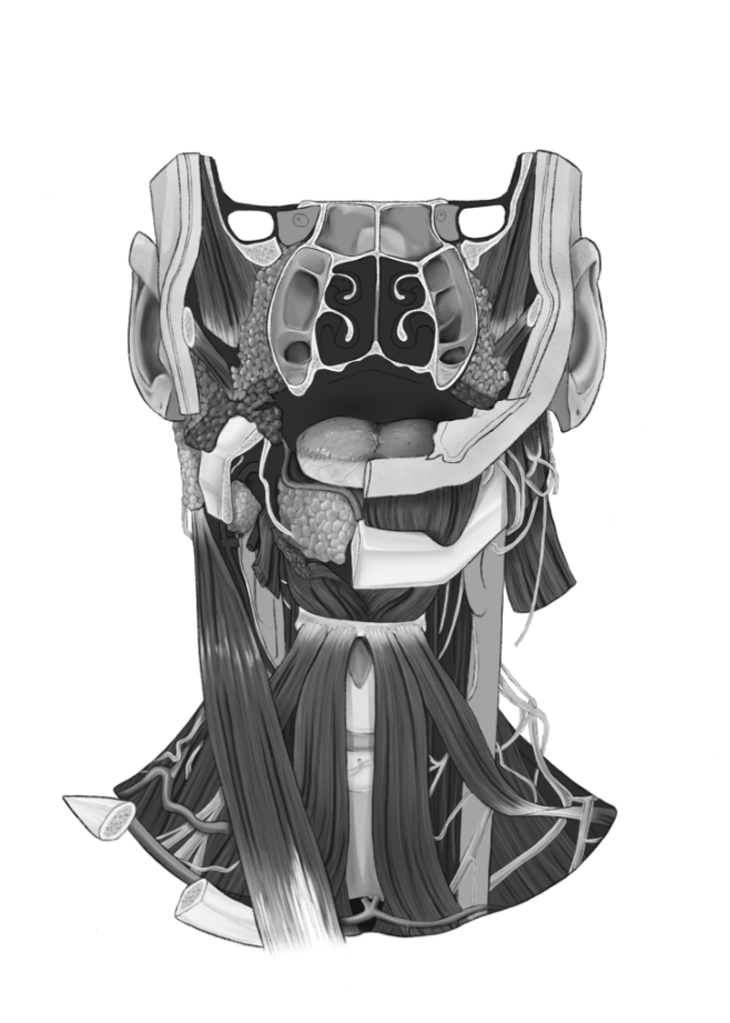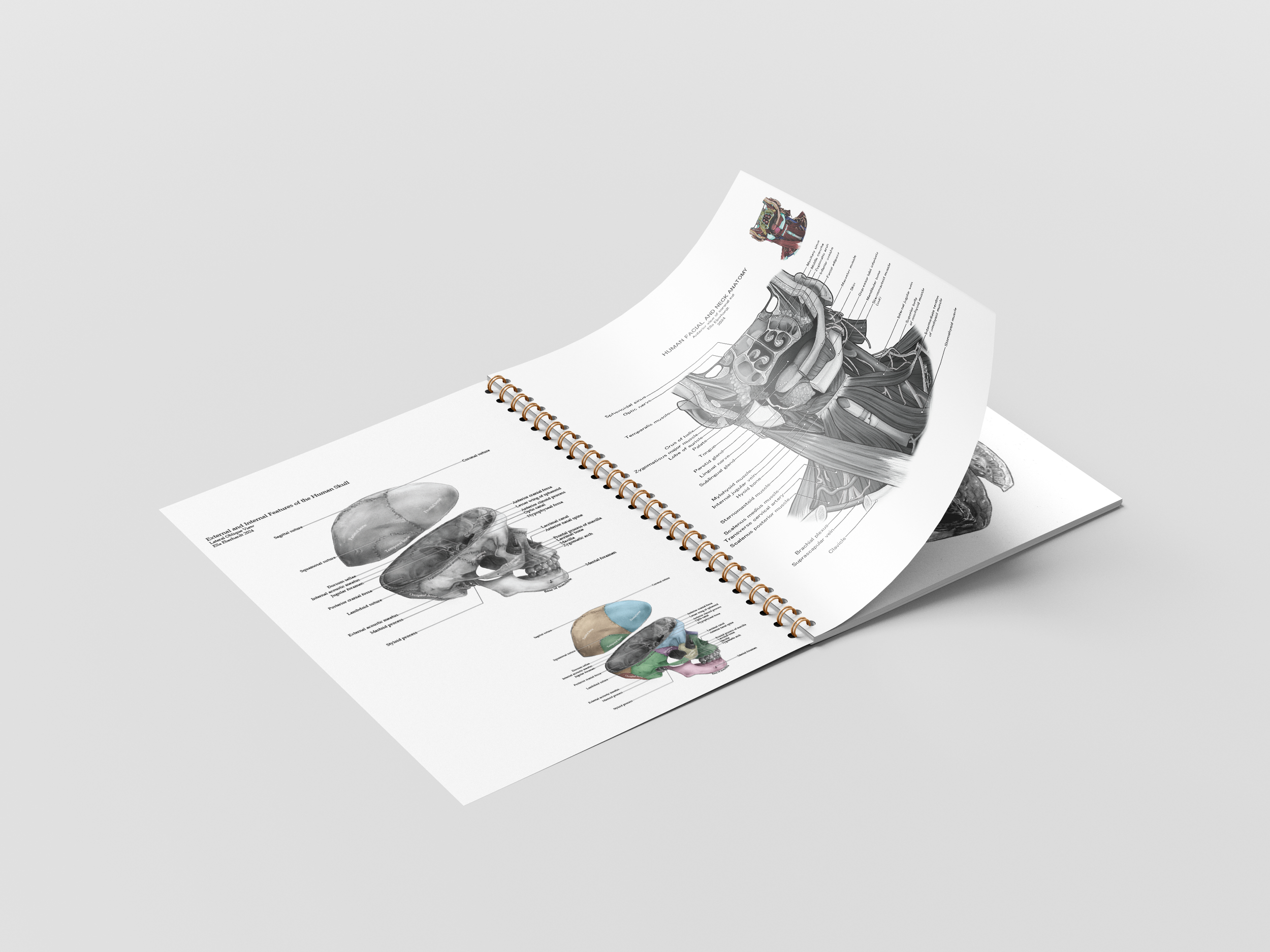
Human Facial and Neck Anatomy
This project focused on the creation of an anatomical diagram from prosected specimens located at Grants Museum on the University of Toronto campus. As these are prosections, part of the goal was to give back life to the specimens muscles and vessels.
Clients
Dave Mazierski (Professor, University of Toronto)
Kristy Cheung (Content expert)
Date
December 2024
Role
Research, Content Development, Visual Development, Layout
Format
1-page anatomical illustration for print
Audience
Anatomy students
Tools
Adobe Photoshop & Procreate
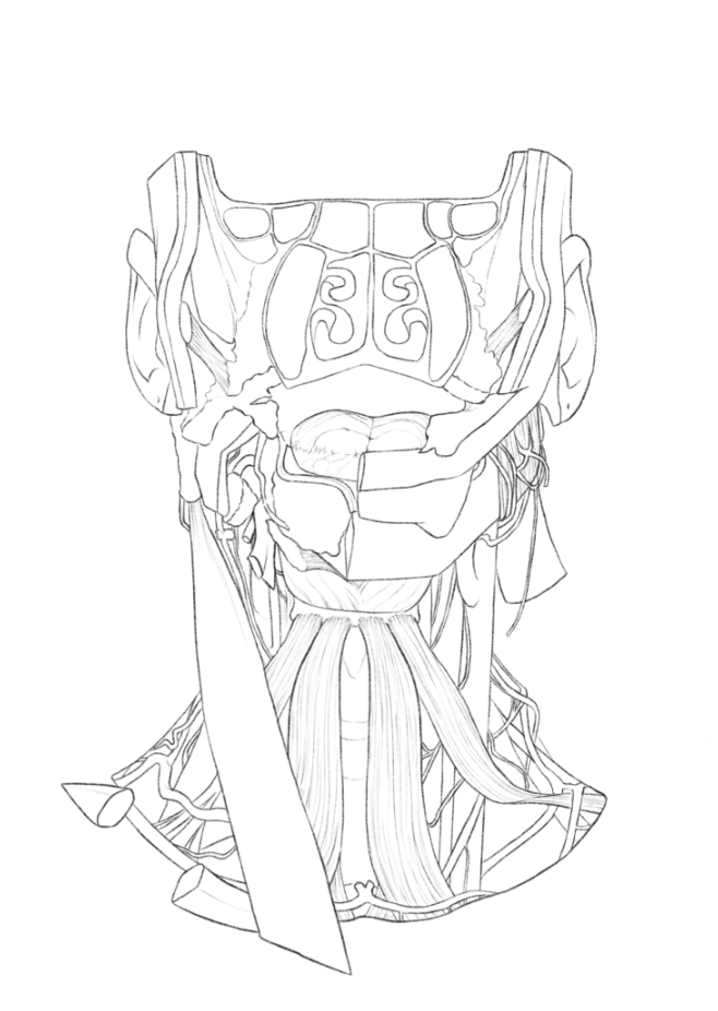
Initial Sketching
The initial sketches for this piece were done sitting in front of the specimen in Grants Museum.
Research
I conducted research on the human facial and neck anatomy looking through several textbooks and consulting with the anatomy department at the University of Toronto.
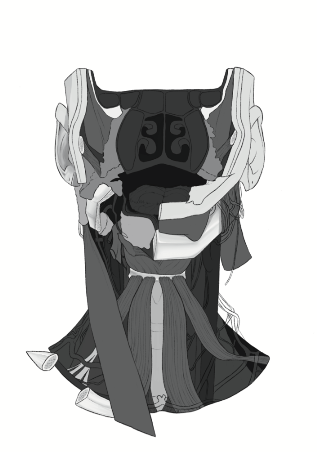
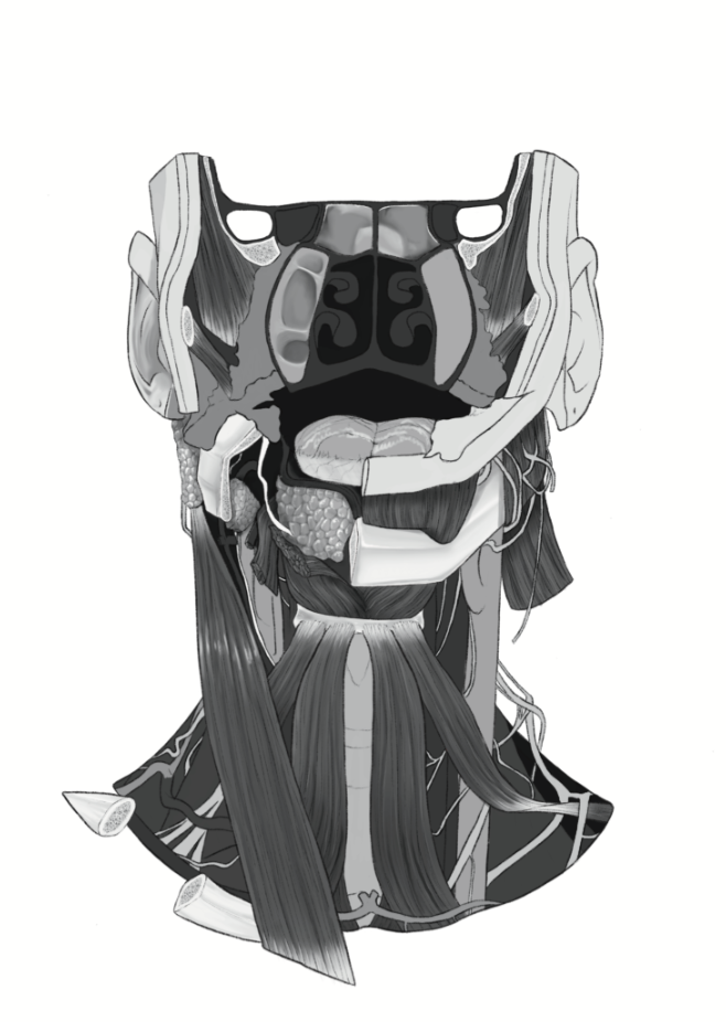
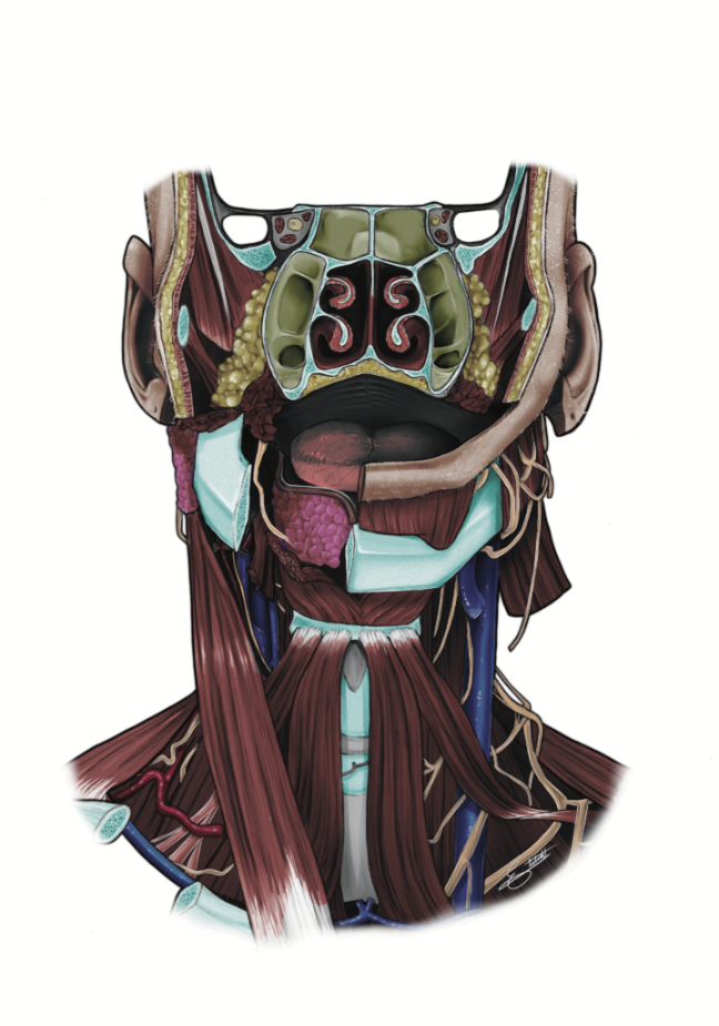
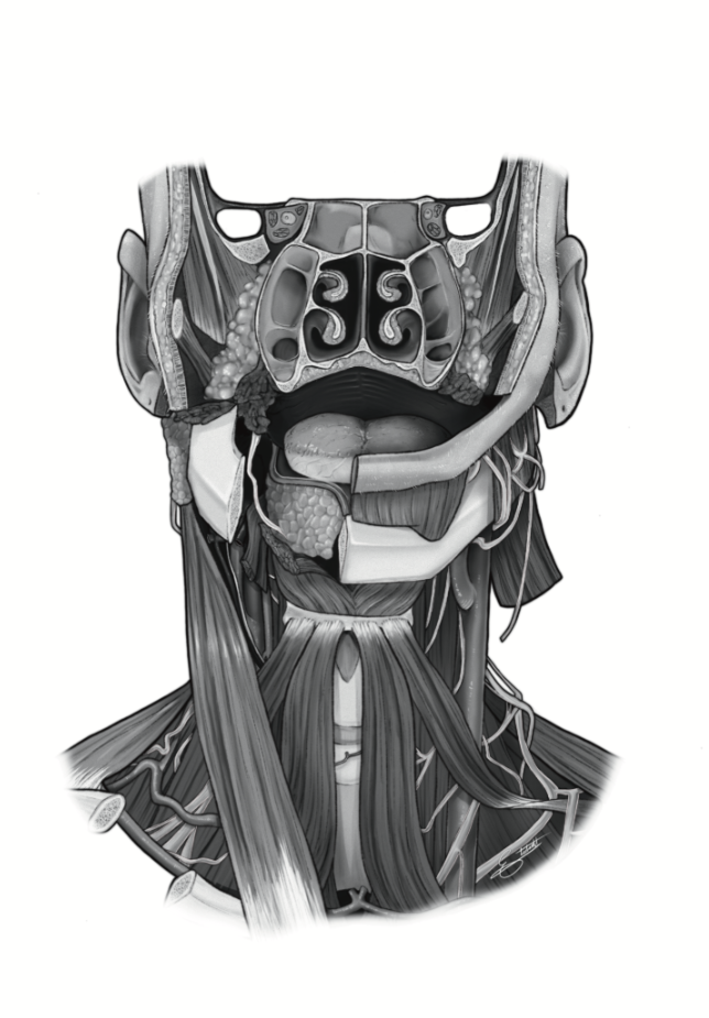
Rendering
This project went through several stages of rendering where I first laid out the main major components, grouping with greyscale before moving in and adding detail and texture. I also created a colourized version that could serve the learner to distinguish between different structures such as bone vs muscle.
References
Agur, Anne M., et al. Essential clinical anatomy. 6th ed., Wolters Kluwer, 2019.
Atlas Anatomique Sandoz, Laboratoires Sandoz, 1973, pp. 7-8.
Netter, Frank H. Atlas of Human Anatomy, 8th ed., Ciba Pharmaceuticals Division - Geigy Corporation, 1995, pp. 26-48, ISBN 0-914168-19-3.
Pascoe, M. Atlas of Human Anatomy, KenHub GmHB, 2023, pp. 446, 460 & 484, ISBN: 978-3-96298-318-5.
Standring, S. Grays Anatomy - The Anatomical Basis of Clinical Practice, 40th ed., Churchill
Livingstone Elsevier Limited, 2008, fig. 30.31 & fig. 32.10, ISBN: 978-0-443-06684-9.